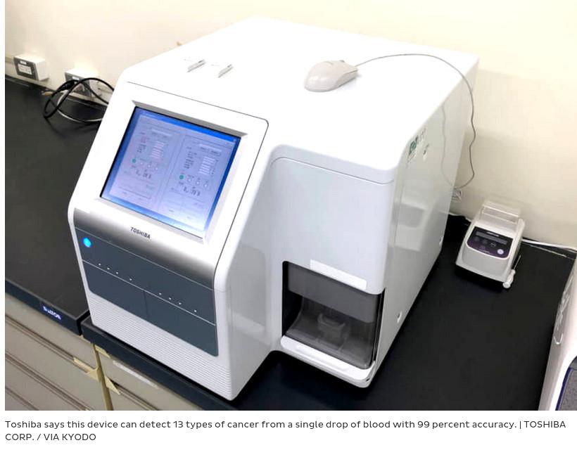
“in one drop of blood 13 types of cancers can be diagnosed early”
those test should be run on the whole population on a yearly basis.
“Early diagnosis and early treatment are the most effective methods to improve prognosis.”
https://www.sciencedirect.com/science/article/pii/S2405844021020223
Kyodo, JIJI
“Toshiba Corp. has developed technology to detect 13 types of cancer from a single drop of blood with 99 percent accuracy, the company announced Monday.
Toshiba developed the diagnosis method together with the National Cancer Center Research Institute and Tokyo Medical University, and hopes to commercialize it in “several years” after starting a trial next year.
The method could be used to treat cancer in its early stage, it said.
The method is designed to examine the types and concentration of microRNA molecules secreted in blood from cancer cells.
Toray Industries Inc. and other companies have also developed technologies to diagnose cancer using microRNA molecules from a blood sample.
“Compared to other companies’ methods, we have an edge in the degree of accuracy in cancer detection,
the time required for detection and the cost,” Koji Hashimoto, chief research scientist at Toshiba’s Frontier Research Laboratory, told a press briefing.
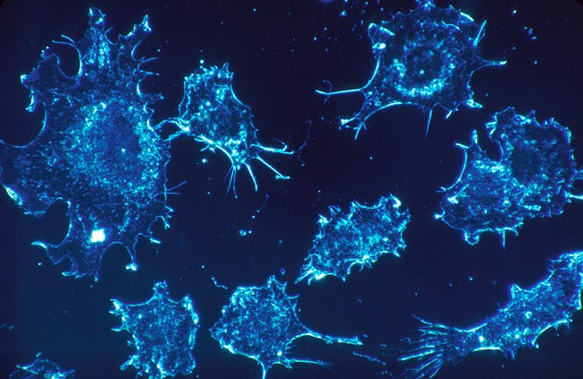
cancer cells blood Cancer cells at a magnification of 500x https://www.inverse.com/article/61214-blood-test-cancer-diagnose-research
Tracking the Sox10 gene Geoffrey Wahl, Christopher Dravis, NCI Salk Institute mobilecancerfactories1512645258606
Treating triple negative breast cancer Wei Qian, National Cancer Institute treatingtriple-negativebreastcancer1512645189680
Where tumor cells enter the bloodstream Elena Deryugina and William Kiosses, Scripps Research Institute wheretumorcellsenterthebloodstream1512645139742

B0003655 Blood vessel in a melanoma, SEM
Credit: S. Gschmeissner, K. Hodivala-Dilke & M. Stone. Wellcome Images
library@wellcome.ac.uk
http://wellcomeimages.org
Colour-enhanced, freeze-fracture scanning electron micrograph of a blood vessel that has grown into a melanoma and is providing nourishment to it. Numerous red blood cells (coloured red) and three white blood cells (coloured yellow) can be seen within the blood vessel (coloured yellow/green). This is surrounded by the melanoma (coloured brown) which is from mouse skin. Horizontal width of image is approximately 230 micrometres.
Scanning electron micrograph
Published: –
Copyrighted work available under Creative Commons Attribution only licence CC BY 4.0 http://creativecommons.org/licenses/by/4.0/
Rumela Chakrabarti, Satrajit Sinha, Yibin Kang, NIGMS Biomedical Beat adevelopingmousemammarygland1512644982157
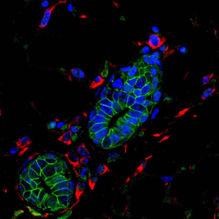
Dr Orli Yogev, Division of Clinical Studies, Institute of Cancer Research 3dconfocalimageofneuroblastomabonemarrowmetastaticneutrospheres1512644914483
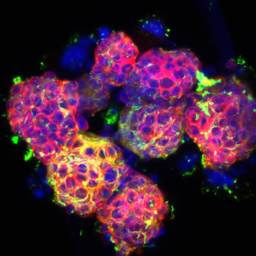
Wei Qian, National Cancer Institute Univ. of Pittsburg Cancer Institute polyploidgiantcancercel1512644861245
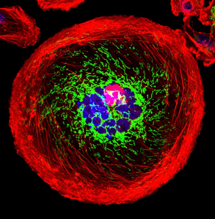
Eric Snyder, National Cancer Institute Huntsman Cancer Institute at the Univ. of Utah kras-drivenlungcancer1512644713615
Picture of Cancer Cell – Thomas Deerinck, National Center for Microscopy and Imaging Research, La Jolla, CA
The test will be used to detect gastric, esophageal, lung, liver, biliary tract, pancreatic, bowel, ovarian, prostate, bladder and breast cancers as well as sarcoma and glioma.
Toshiba has developed a chip and a small device that can conduct the diagnosis in less than two hours. A blood test using it is expected to cost ¥20,000 (164€
/ 182$) or less, it said.
It is possible that the device will be used in health checkups, the company said.
In its five-year business strategy from April 2019, Toshiba has positioned medical businesses, including genome analysis and cell diagnosis, among key growth pillars along with automation, batteries and digital solutions using artificial intelligence.”
https://www.japantimes.co.jp/news/2019/11/25/business/corporate-business/toshiba-test-cancer-blood/
https://www.democracynow.org/2019/12/23/the_first_cell_dr_azra_raza_on
Amy Goodman: “no major improvement in cancer treatment since 50 years”

“diagnose early and nip them in the but”
https://www.amazon.de/First-Cell-Human-Pursuing-Cancer/dp/1541699521/
https://en.wikipedia.org/wiki/Azra_Raza
website: https://azraraza.com/
more videos: https://azraraza.com/video-category/
liked this article?
- only together we can create a truly free world
- plz support dwaves to keep it up & running!
- (yes the info on the internet is (mostly) free but beer is still not free (still have to work on that))
- really really hate advertisement
- contribute: whenever a solution was found, blog about it for others to find!
- talk about, recommend & link to this blog and articles
- thanks to all who contribute!